University of Bielefeld, Medical School EWL.

Live Cell Imaging
Live Cell Imaging of immortalized osteosarcoma cell line using the Leica Stellaris 8 TauSTED
June 2023

The limits of light microscopy have been overcome by breakthrough technologies in the last decade. Several research groups around the world have developed new methods to improve the resolution for the identification of cellular events, particularly in the area of live cell imaging. However, super-resolution microscopes are a very expensive and complex type of laboratory equipment that not every lab can afford.
In addition, it can take several months or years to master the operation of the instrument, making it even more difficult to utilize. This report describes the use of a Cellbox™ Shipper Ground to transport U-2OS cells from a laboratory at the Medical Faculty OWL, University Bielefeld to a demonstration facility in Mannheim.
The Project
The advantage of live cell imaging is the visualization of transient and dynamic cellular processes avoiding fixation artifacts. This technology is especially critical for the study of signaling pathways and molecular interactions. In this project, U-2OS cells (an immortalized osteosarcoma cell line) stably expressing different genetic constructs were seeded on day one at 5x10³ cells/ml in ibidi 8-well μ-slides at 300 μl per well and in 35 mm dishes (MatTek) at 2 ml per dish using DMEM without phenol red supplemented with 10% fetal calf serum and antibiotics.
Cells were grown for two days. The Cellbox™ was packed with confluent U-2OS cells at 37°C and 5% CO2 on day three. Cells were kept in the Cellbox™ during the cab ride between the University of Bielefeld and the Bielefeld central station, the subsequent train ride from Bielefeld central station to Mannheim central station, and the cab ride between Mannheim central station and a local hotel for an overnight stay. On day four, the Cellbox™ was transported from the local hotel in Mannheim to the Leica demonstration center.
The Outcome
All planned imaging experiments were successful and the cells were examined under the microscope as confluent, viable and with vibrant activity.
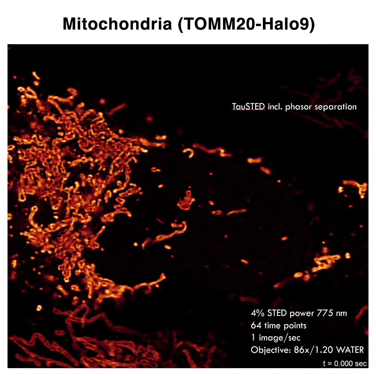
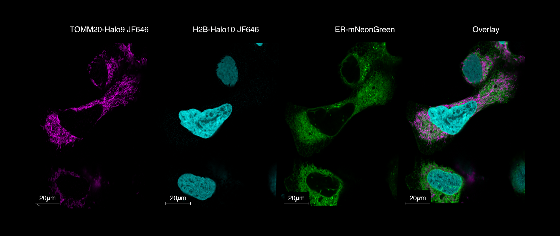
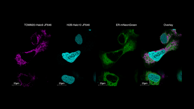
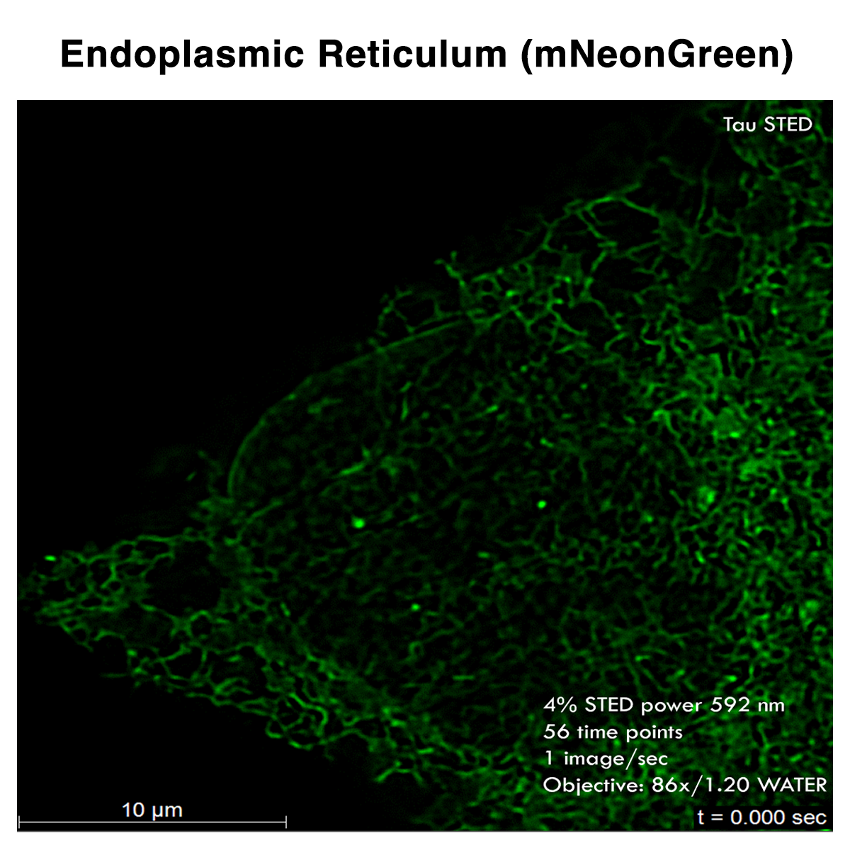
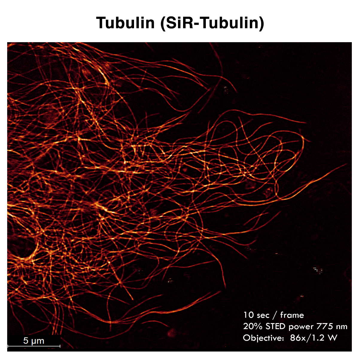
U-2OS cells expressing differentially labeled proteins were examined by the superresolution approach STED, and its FLIMsupported variant TauSTED, to visualize membranous structures and protein interactions, such as the endoplasmic reticulum (ER-mNeonGreen), peroxisomes (YFP-SKL), mitochondrial outer membrane (TOMM20-Halo9), histones (H2B-Halo10) and tubulin (SiR-Tubulin), which showed high viability and structural integrity. In addition, ER, tubulin, and mitochondria dynamics as well as mitochondrial fusion and fission processes (Drp1-SNAP + SiR-SNAP) could be observed, which is a proven success for the Cellbox™ overnight transport from University Bielefeld to the Leica demonstration site in Mannheim.
